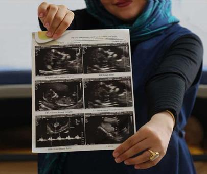
In the imaging department, the ultrasound of the first trimester of pregnancy is performed from the beginning of the 11th week of pregnancy (11W+0D) to the end of the 13th week of pregnancy (13W+6D), when the fetal length (CRL) is in the range of 45 to 84 mm, and it is the right time for Screening for common chromosomal abnormalities in the first trimester of pregnancy. During this period, a pregnant person can be screened for chromosomal abnormalities in the first trimester.
The obstetrician calculates the due date as 280 days or 40 weeks after the first day of the last menstrual period (LMP). For women who have a regular 28-day menstrual cycle, this method is almost accurate, and they can calculate their gestational age with LMP and perform screening at the right time, but when the cycles are irregular, many miscalculations occur. At this time, or if they have forgotten their LMP, it is necessary to have a regular ultrasound to control the CRL (fetal length) and estimate the gestational age. The best ultrasound time to determine the gestational age is to perform an ultrasound at 8-9 weeks.
Considering that the common defects and syndromes under investigation including Down's syndrome (trisomy 21), Edward's syndrome (trisomy 18), and Pato's syndrome (trisomy 13) are also seen in families that have no history, so it is recommended that Let all women at any age perform screening tests for chromosomal abnormalities and necessary ultrasounds. On the other hand, in the ultrasound of the first trimester, the length of the fetus (CRL), which determines the gestational age, is measured accurately and according to the standards of the International Fetus Association (FMF), which determines the age and growth of the fetus in the design of pregnancy. It is of fundamental importance

